The exact nature of long COVID is still coming to light, but we just got some of the best evidence yet that this debilitating condition stems from a brain injury.
Using high-resolution scanners, researchers at the Universities of Cambridge and Oxford have shown microscopic, structural abnormalities in the brainstems of those recovering from COVID-19.
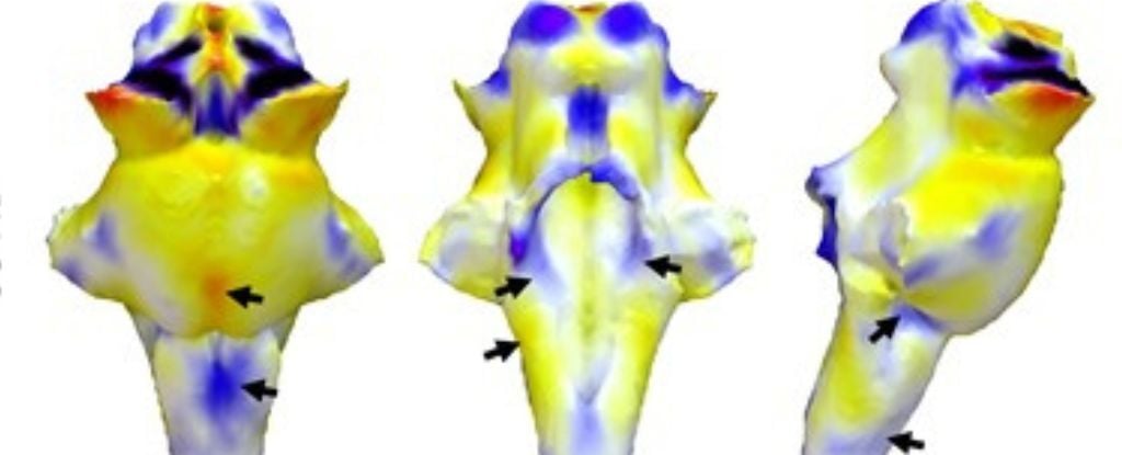
Signs of brain inflammation were present up to 18 months after first contracting the SARS-CoV-2 virus.
[…]
In living brains of those with long COVID, however, conventional MRI studies have shown no structural abnormalities in the brainstem.
Do these people not proof-read their own articles?
Normal hospital-type MRI scanners can’t see inside the brain with the kind of chemical and physical detail we need. But with 7T (7 Tesla) scanners, we can now measure these details
Not the best article, but I think what they are trying to say over multiple paragraphs is that new higher resolution MRI machines can see the damage that normal lower resolution MRI can’t see
FTA: Signs of ongoing inflammation in the brainstem, something that is seen in people with traumatic brain injury and people with chronic fatigue syndrome.
so this new study is saying they can see on a small enough scale to find that scientifically consistent and significant inflammation in the brainstem directly linked to covid?
Precisely. They had previously been unable to see this in living patients due to limitations of typical MRI machines but had found signs in the deceased. The major change is that, using a more powerful (7 Tesla) MRI machine, they were able to see these same symptoms in living patients for the first time.
yes, I can guess that explanation when trying to figure out the seeming contradiction. I don’t read scientific articles to end up guessing because the author can’t string together a well structured text. :)
I have to choose what to spend my time on. If an article contradicts itself that obviously after I spent 2-5 minutes reading, I’ll go look for more intelligent texts.
It’s not contradicting itself though. Your first quoted statement says “using high-resolution scanners”. The last one says “conventional MRI studies”. The methodology is what is different.
Let me break it down so you see the point I was making - in case the bold wasn’t enough:
Using high-resolution scanners, researchers at the Universities of Cambridge and Oxford have shown microscopic, structural abnormalities in the brainstems of those recovering from COVID-19.
Signs of brain inflammation were present up to 18 months after first contracting the SARS-CoV-2 virus.
Here, they refer to people recovering from COVID-19, thus clearly indicate that patients are alive.
[…]
In living brains of those with long COVID, however, conventional MRI studies have shown no structural abnormalities in the brainstem.
This paragraph immediately follows one that talks about autopsy(!) results, and here, they start a sentence with “in living brains […], however”, setting the sentence up as a contradiction to the previous one, with an emphasis on the word living in the article itself.
Here’s an example how the sentence should be written to not seemingly cause a contradiction / misdirect the reader:
However, previous studies conducted with conventional MRI had shown no structural abnormalities in the brainstem in living brains.
They put emphasis on the change in observation from autopsy to living brains, linking this paragraph more strongly to the preceeding one, when they should have put emphasis on the conventional studies, building the context for the subsequent paragraph.
I don’t really understand what the difference between the fixed and og version is.
The fixed version is slightly is better, I agree, but I wouldn’t call it a necessary fix.
You are judging a field specialist(s) on basically their communication skills. We can’t all structure sentences well, brainholes work differently, wording thoughts is hard.
It’s a bit like bitching how a perfectly working shovel isn’t ornate enough since if it was it would have been more pleasant to work with - it’s prob true but it’s such a low gain ‘nice-to-have’ feature people generally don’t bother with it.
Imho narratives need to be pleasant and/or artistic, eg I expect a novel to be written good (tho absolutely not a huge point for me), I don’t expect that from a game theory book, I expect it to be correct. (Another example might be how stupidity convoluted laws/contracts/t&a are written.)
What I kinda demand (only slightly irrationally or at least to an impractical extend) is that the subject is conveyed in an exact manner. I expect exact communicating overall.
And the og text you quoted is exact.
Oh … and at both ends it’s literacy - their (+to some extend whoever proofread it) literacy levels shows how eloquently they conveyed their data & thoughts, your literacy level shows how you though they are contradicting themselves.
Oh, a journalist, then bitch on :D (no /s, after all, random bitching is a fair part of lemmy).
But I think people replied to your og message because of the (mis?)use of the word “contradictory”, not because of the bitching as such.
(And Im mostly here bcs I like to understand myself, like what triggered you sceptically & how the same thing played in my mind)
because of the ~~(mis?)~~use of the word “contradictory”,
I used that only in my second comment, after the first person got flustered :) Go up two more in the comment chain and you’ll see my original comment.
Although, I stand by the second comment as well - the article is contradicting itself.
If I say
The square root of -1 is i
[…]
The square root of -1 is not defined.
[…]
Only to THEN go on to explain what imaginary numbers are, then I have still contradicted myself :)

Do these people not proof-read their own articles?
Normal hospital-type MRI scanners can’t see inside the brain with the kind of chemical and physical detail we need. But with 7T (7 Tesla) scanners, we can now measure these details
Not the best article, but I think what they are trying to say over multiple paragraphs is that new higher resolution MRI machines can see the damage that normal lower resolution MRI can’t see
what type of abnormalities are they seeing specifically?
FTA: Signs of ongoing inflammation in the brainstem, something that is seen in people with traumatic brain injury and people with chronic fatigue syndrome.
oh, interesting.
so this new study is saying they can see on a small enough scale to find that scientifically consistent and significant inflammation in the brainstem directly linked to covid?
Precisely. They had previously been unable to see this in living patients due to limitations of typical MRI machines but had found signs in the deceased. The major change is that, using a more powerful (7 Tesla) MRI machine, they were able to see these same symptoms in living patients for the first time.
okay, got it. thanks.
that is a heck of a development, I now understand the cause for the hullabaloo.
You’re very welcome! Glad that I could help.
Removed by mod
deleted by creator
deleted by creator
The abnormalities are only visible with a 7T scanner, and not conventional MRIs.
yes, I can guess that explanation when trying to figure out the seeming contradiction. I don’t read scientific articles to end up guessing because the author can’t string together a well structured text. :)
No guesswork was needed, only a modicum of reading comprehension.
Dude read the rest of the article
I have to choose what to spend my time on. If an article contradicts itself that obviously after I spent 2-5 minutes reading, I’ll go look for more intelligent texts.
It’s not contradicting itself though. Your first quoted statement says “using high-resolution scanners”. The last one says “conventional MRI studies”. The methodology is what is different.
Let me break it down so you see the point I was making - in case the bold wasn’t enough:
Here, they refer to people recovering from COVID-19, thus clearly indicate that patients are alive.
This paragraph immediately follows one that talks about autopsy(!) results, and here, they start a sentence with “in living brains […], however”, setting the sentence up as a contradiction to the previous one, with an emphasis on the word living in the article itself.
Here’s an example how the sentence should be written to not seemingly cause a contradiction / misdirect the reader:
They put emphasis on the change in observation from autopsy to living brains, linking this paragraph more strongly to the preceeding one, when they should have put emphasis on the conventional studies, building the context for the subsequent paragraph.
I don’t really understand what the difference between the fixed and og version is.
The fixed version is slightly is better, I agree, but I wouldn’t call it a necessary fix.
You are judging a field specialist(s) on basically their communication skills. We can’t all structure sentences well, brainholes work differently, wording thoughts is hard.
It’s a bit like bitching how a perfectly working shovel isn’t ornate enough since if it was it would have been more pleasant to work with - it’s prob true but it’s such a low gain ‘nice-to-have’ feature people generally don’t bother with it.
Imho narratives need to be pleasant and/or artistic, eg I expect a novel to be written good (tho absolutely not a huge point for me), I don’t expect that from a game theory book, I expect it to be correct. (Another example might be how stupidity convoluted laws/contracts/t&a are written.)
What I kinda demand (only slightly irrationally or at least to an impractical extend) is that the subject is conveyed in an exact manner. I expect exact communicating overall.
And the og text you quoted is exact.
Oh … and at both ends it’s literacy - their (+to some extend whoever proofread it) literacy levels shows how eloquently they conveyed their data & thoughts, your literacy level shows how you though they are contradicting themselves.
Or am I?
Also, it’s not my fault that people got all flustered about me simply pointing out that poor phrasing with “do they even proofread?”
Edit: goat -> got
Oh, a journalist, then bitch on :D (no /s, after all, random bitching is a fair part of lemmy).
But I think people replied to your og message because of the (mis?)use of the word “contradictory”, not because of the bitching as such.
(And Im mostly here bcs I like to understand myself, like what triggered you sceptically & how the same thing played in my mind)
I used that only in my second comment, after the first person got flustered :) Go up two more in the comment chain and you’ll see my original comment. Although, I stand by the second comment as well - the article is contradicting itself.
If I say
The square root of -1 is i […] The square root of -1 is not defined. […]
Only to THEN go on to explain what imaginary numbers are, then I have still contradicted myself :)