From Blackland Prairie Raptor Center
Let’s walk through some parts of an examination of a bird.
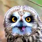
An Eastern Screech Owl was brought to our Raptor Clinic because it was lethargic, weak, and dehydrated. First, a cloth hood is placed over the head. Covering a bird’s head helps keep it calm. Our veterinarian performed a physical body check of the owl – including ears, feet, and eyes. (Above)


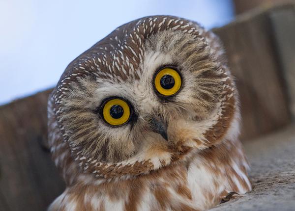
Our vet put drops in the eyes to help see any possible eye conditions that may need attention. When applied, the eye drops appear yellow and fluoresces green under blue light, allowing our vet to visualize damaged areas on the cornea more clearly.


The owl was taken to our x-ray room. This patient was very inactive, which made it easy to arrange it on the x- ray table. Wings are spread out and the feet and tail are extended and then taped down. The x-ray is taken and can quickly be viewed on our computer monitor. Our vet reviews the x-ray for fractured or broken bones; issues with organs, embedded foreign objects, etc. Fortunately, the x-rays looked good for this little owl. The owl was taken back to the exam room.


Medications (such as anti-inflammatory) were given orally through the mouth and hydration liquids were given through an IV in its leg.
The owl was then placed in our Intensive Care Unit.
Initially, the owl stayed very still, quiet, eyes were closed, and ate a little food. The second day, it was standing on its food tray – good sign that it’s moving and probably looking for food. On the sixth day, the owl’s eyes were open, it was more alert, and showed defensive signs of clacking its beak. The owl continued to improve over the next few days and was moved from ICU to an outdoor mew. The owl can continue to get healthy and build up its strength by flying around in the mew.
Distraction Tabs