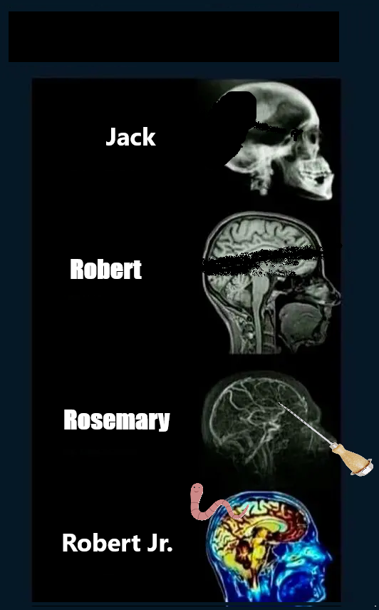
Holy shit that is so wrong, and I am here for it.
Jesus Fucking Krist
Removed by mod
Removed by mod
Removed by mod
Turn left on to Kennedy blvd.
Having to look up MRA. It’s just MRI (NMR) but with contrast that goes specifically to the brain (maybe carb-based)?
Magnetic Resonance Angiogram. Yeah it’s an MRI. I guess they’re processing it somehow to enhance bloodflow like FMRI. Seems like it doesn’t require contrast.
Radiologist here. 👋🏻
You are right it does not require contrast. The scan uses a special technique to reduce signal from tissue that isn’t moving so only things that are moving produce high signal and show up “bright.” Since blood is the only thing really moving in the brain - voila!
Neat, I was thinking it was something like motion amplification. I guess the lungs would mess with imaging of the torso, or can you pick the motion frequency to isolate bloodflow but ignore respiration?
The lung motion is be a problem but it is still done in some situations and the radiologist just tries their best with any motion artifacts. Since the blood vessels are so big in the chest it kind of works out, but it turns out better with contrast.
I see. I started reading this article which describes a whole host of different techniques. It really seems to be exploiting the physics of NMR to get this enhancement (or deficit), along with the image processing. It’s really interesting but I’d need a while to get my head around it.
Malady of the brain

Is it really an x-ray of the brain? Surely that’s just of the skull.
Technically it’s both. It’s just that the bones stand out much more than the soft squishy insides.
All of them are XYZ of the head,
Nice
There are brain x-rays called pneumoencephalographs where they replace the cerebrospinal fluid with air, x-ray, and then put the fluid back. Pneumoencephalography and angiography were really the only neuroimaging techniques available for most of the 20th century, so it was pretty common.
Does the brain belong to Vince McMahon?
It’s the perfect antimeme
I love it!


